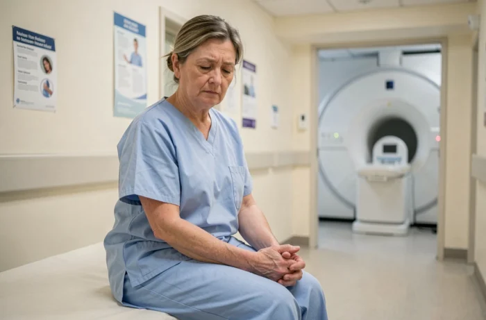
The distinction between standard closed MRI, wide-bore MRI, and open MRI directly affects the feasibility of the examination for a claustrophobic patient. Confusing these three configurations means directing a patient to a machine that will not solve their problem or imposing unnecessary sedation.
Wide-bore MRI versus open MRI: two technical responses to different levels of anxiety
The standard closed MRI offers a tunnel with a diameter of about 60 cm. The wide-bore MRI increases this diameter to 70 cm, which is sufficient to significantly reduce the rate of interrupted exams in patients with moderate anxiety. For a confirmed phobia, this 10 cm difference does not change the situation: the patient remains in a tunnel, and the feeling of confinement persists.
The open MRI eliminates the tunnel. The patient lies between two horizontal plates, with complete lateral clearance. For patients who are truly phobic, this is often the only configuration that allows the examination to be performed without sedation or general anesthesia.
In practice, we observe a logical progression: prior discussion and feet-first positioning, then wide-bore if anxiety persists, then open MRI, then light sedation, and finally anesthesia as a last resort. Feedback from patients available in the reviews on open MRI for claustrophobes confirms that this step-by-step approach prevents the majority of anxious patients from resorting to medication sedation.
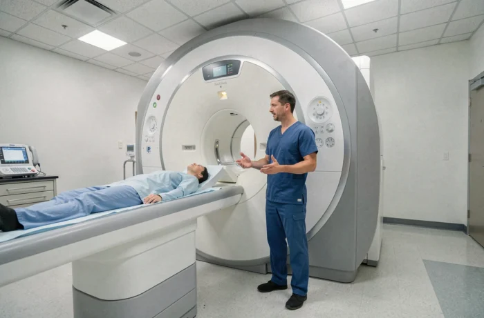
Image quality in open MRI: the real compromise on diagnosis
One point that popular articles consistently overlook: open MRI operates at a lower magnetic field than closed machines. Most open MRIs operate between 0.2 T and 0.7 T, compared to 1.5 T or 3 T for closed machines.
This difference in field has direct consequences on the signal-to-noise ratio and spatial resolution. For a brain MRI searching for small lesions, or for a detailed joint assessment of ligaments, the loss of resolution can compromise the diagnosis.
Indications where open MRI remains reliable
- Standard spinal assessment (herniated disc, narrow lumbar canal): the resolution is sufficient in the vast majority of cases
- MRI of large joints (knee, shoulder) for meniscal lesions or significant tendon ruptures
- Follow-up of already identified pathologies, where images are compared to a previous reference examination
Indications where the compromise becomes problematic
- Fine brain exploration (multiple sclerosis, search for micro-vascular lesions)
- Liver assessment with dynamic sequences after gadolinium injection
- Functional cardiac imaging, which requires a high field and fast sequences
Accepting an open MRI for claustrophobia may degrade the diagnostic value in these specific indications. We recommend discussing this point with the referring radiologist before choosing the type of machine, rather than discovering afterward that a second examination in a closed MRI will be necessary.
Anxiolytic premedication before MRI: what patients really report
The prescription of a mild anxiolytic before the examination remains the most frequently proposed first-line solution. In practice, feedback from anxious patients strongly nuances the effectiveness of this approach.
A tablet of hydroxyzine or a low-dose benzodiazepine taken an hour before the examination alleviates anticipatory anxiety. It does not eliminate the phobic reaction triggered by entering the tunnel. Some patients describe a dissociation between a state of drowsiness and intact panic, which paradoxically makes the experience more destabilizing.
Premedication works mainly on moderate anxiety, not on established phobia. For the latter, monitored intravenous conscious sedation, performed in a hospital environment, offers more reliable control. However, it requires different organization: mandatory companion, post-examination monitoring, inability to drive.
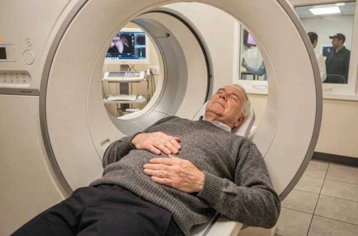
Non-drug techniques tested by patients: beyond abdominal breathing
Abdominal breathing and music through headphones are mentioned everywhere. Their usefulness is real but limited in patients whose phobia exceeds simple discomfort. Three less documented strategies emerge from patient testimonies.
Feet-first positioning in the tunnel radically changes the perception of confinement for MRIs of the spine, pelvis, or lower limbs. The head remains outside or at the entrance of the tunnel, and the feeling of confinement decreases significantly. This positioning is not possible for brain MRIs, but it is underutilized for other indications.
Conversational hypnosis, practiced by some trained technicians, acts during the examination itself. The caregiver maintains a calibrated verbal exchange via the intercom, directing the patient’s attention towards sensory representations incompatible with panic. Some radiology centers now offer this training to their teams.
Prior familiarization with the examination room, outside of any appointment, allows the patient to touch the machine, lie on the table without starting a sequence, and test the call button. This desensitization through gradual exposure, borrowed from behavioral therapy protocols, is rarely offered due to a lack of available slots.
The choice between open MRI, wide-bore with adapted positioning, or closed MRI under premedication depends on the patient’s actual anxiety level and the diagnostic requirements of the prescribed examination. A prior phone exchange with the radiology department, describing their previous reactions to confined spaces, helps guide towards the most suitable configuration without improvising on the day of the examination.